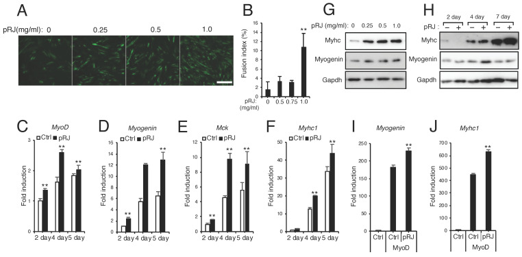
Figure 5.
pRJ stimulates myoblast differentiation. (A,B) C2C12 cells were treated with myogenic medium supplemented with pRJ at 0, 0.25, 0.5, or 1.0 mg/mL for 6 days. Cells were then stained with anti-myosin heavy chain antibody (A). Fusion index was quantified as the number of nuclei (at least three) within myotubes divided by the total number of nuclei (B). (C–F) Cells were treated with myogenic medium together with or without 1 mg/ml pRJ for 2, 4, or 5 days. mRNA levels of the indicated myogenic markers were determined by qPCR. (G and H) C2C12 cells were treated with myogenic medium supplemented with pRJ at 0, 0.25, 0.5, or 1.0 mg/mL for 4 days (G) or treated with 1 mg/mL pRJ for 2, 4, or 7 days (H). Western blots showing protein levels of myosin heavy chain (Myhc), Myogenin, or Gapdh (G and H). (I and J) C3H10T1/2 cells were transfected with or without MyoD and then treated with or without 1mg/mL pRJ. mRNA levels of Myogenin or Myhc1 were determined on day 2 by qPCR. Representative images are shown. Scale = 100 μm (A, G, and H). Data are mean ± SD (n = 4). ∗∗p < 0.01, ∗p < 0.05, versus control (Ctrl).

