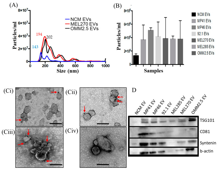
Figure 1.
Characterization of extracellular vesicles (EVs) derived from normal choroidal melanocytes (NCMs) and Uveal Melanoma (UM) cells. (A,B) Nanoparticle tracing analysis NTA of EVs derived MEL270, metastatic UM cells (OMM2.5) and NCMs. (B) NTA data showing concentrations of EVs from different cell sources. Data are expressed as mean ± SD (n = 3). (C) Representative micrographs of immunoGold-TEM on MP46-EVs (Ci–Cii) 92.1-EVs (Ciii) and NCM-EVs (Civ) labelled with a cocktail of antibodies against CD81 (Ci), TSG101 (Cii) and CD63 (Ciii) (red arrows). Scale bars 200 nm. (D) Proteins isolated from EVs derived from different cell sources were analyzed by Western blot for the expression of specific exosome markers.

