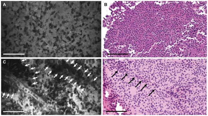
Figure 2.
Photomicrographs of a pituitary adenoma. (A) Confocal laser endomicroscopy (CLE) image (see Supplementary Video S1) and (B) hematoxylin and eosin (H&E) stain of the same tumor showing sheets of uniform nonlobulated cells with prominent nuclei. (C) CLE image (see Supplementary Video S2) and (D) H&E image showing perivascular sheets of the cells (arrows). Bar = 100 μm. Used with permission from Barrow Neurological Institute, Phoenix, Arizona.

