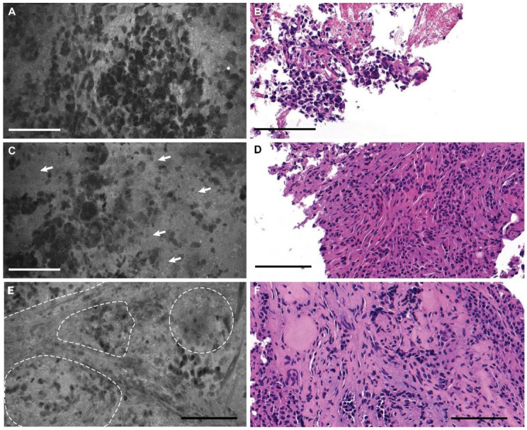
Figure 5.
(A) Confocal laser endomicroscopy (CLE) image that was nondiagnostic because the biopsy specimen was acquired too early (<1 min after fluorescein injection); (B) a hematoxylin and eosin (H&E)-stained section from the same specimen. (C) CLE image that was nondiagnostic because the biopsy specimen was acquired too late (>10 min after fluorescein injection), with arrows pointing to the hypercellular areas of tissue with suboptimal contrasting of the cellular outlines; (D) an H&E-stained section from the same specimen. (E) CLE image showing more uniform lobules of pituitary epithelial cells, suggestive of normal pituitary tissue, with dotted lines outlining lobules; (F) H&E-stained section from the same specimen. Bars = 100 μm. Used with permission from Barrow Neurological Institute, Phoenix, Arizona.

