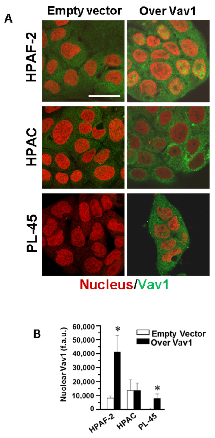
Figure 3.
Confocal analysis of nuclear Vav1 in PDAC-derived cell lines. (A) Representative confocal images of HPAF-2, HPAC, and PL-45 cells transfected with a construct expressing the full-length human Vav1 (Over Vav1) and stained with the antibody against Vav1 (green). DRAQ5 stain was used to counterstain the nucleus (red). Bar = 20 μm. (B) Fluorescence intensity of nuclear Vav1 measured by the ImageJ software on digitized confocal images. F.a.u.: fluorescence arbitrary unit. Error bars indicate ± SD from a triplicate experiment. * p < 0.05.

