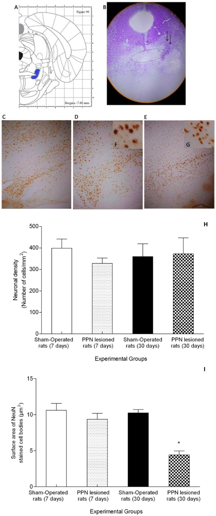
Figure 10.
Morphological study. (A) Schematic diagrams of the sections showing pedunculopontine nucleus (PPN) area (blue color) adjacent to the superior cerebellar peduncle (SCP) in the rat brain adapted from Paxinos and Watson (2007). The diagram corresponds with the antero posterior coordinate used in the present study. (B) Representative photomicrographs of the cresyl violet study showing the location of the PPN lesion (4×). The ellipse represents the SCP bundle in whose distal portion the area corresponding to the pedunculopontine nucleus is located. Little black arrows indicate the lesion area. C-G Representative photomicrographs of the immunohistochemical study for anti-neuronal nuclear protein (NeuN) in nigral coronal sections. (C) Sham-operated rats (10×). (D) Right substantia nigra (seven days post PPN lesion) (10×). (E) Right substantia nigra (30 days post PPN lesion) (10×). (F,G) Magnification (40×) of the image (D,E). (H) Comparison of neuronal density between the right substantia nigra of PPN-lesioned rats (seven and 30 days after lesion) and Sham-operated rat groups (Z = 3.74 p > 0.05). (I) Comparison of surface area between the right substantia nigra of PPN-lesioned rats (seven and 30 days after lesion) and Sham-operated rat groups (Z = 9.77 p < 0.05). * p < 0.05.

