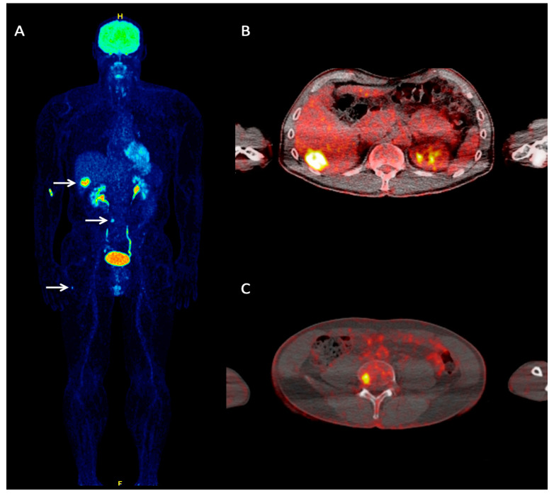
Figure 1.
A 60-year-old Merkel cell carcinoma (MCC) patient referred to our department for staging purposes due to clinical suspicion of hepatic metastases. Whole-body 18F-FDG PET maximum intensity projection (MIP) (A) demonstrated foci of increased tracer uptake in the liver, lumbar spine, and right femur, corresponding to metastases (arrows). Transaxial, fused 18F-FDG PET/CT at the hepatic level (B) shows a focal site of increased tracer uptake in liver segment VI, corresponding to a hepatic metastasis. Transaxial, fused 18F-FDG PET/CT of the lower abdomen (C) shows pathologic tracer accumulation in the fourth lumbar vertebrae, representing an osseous metastasis.

