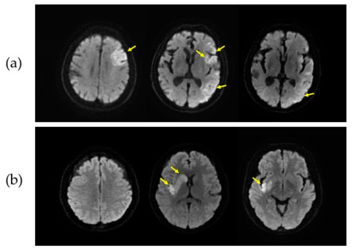
Figure 1.
Representative diffusion-weighted magnetic resonance imaging (DWI) slices from two groups. DWI data from a patient with the Alberta Stroke Program Early Computed Tomographic Score (ASPECTS) of 5 (a) and the ASPECTS of 7 (b) are shown. The slices in the first, second and third column in each patient correspond to the supraganglionic, ganglionic, and infraganglionic levels, respectively. The yellow arrows indicate infarct lesions.

