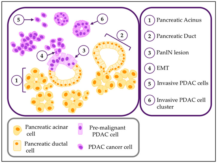
Figure 1.
Normal and PDAC parenchymal cells. Diagram representing the parenchymal cellular components of the normal exocrine pancreas, PanIN pre-malignant lesions and PDAC. Histological features of each includes acini (1), ducts (2), atypic cells in panIN lesions (3), PDAC cells undergoing epithelial-to-mesenchymal transition (4), invasive PDAC migrating as individual cells.

