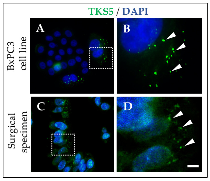
Figure 2.
TKS5-positive invadopodia in a PDAC cell line in culture and in a PDAC archived surgical specimen. (A) BxPC3 cells were stained with a TKS5 antibody and DAPI. (B) Image corresponding to square in A. (C) Sections from an archived paraffin-embedded PDAC surgical specimen stained with a TKS5 antibody and DAPI. (D) Image corresponding to square in C. Arrowheads, invadopodia (B) and invadopodia-like structures (D). Bar, 1 μm in A, C and 0.1 μm (B,D). See also Refs. [48,49,50].

