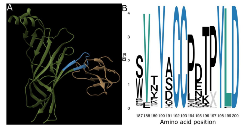
Figure 1.
The ligand-binding domain of the nicotinic acetylcholine receptor (nAChR). (A) Ribbon model of α-bungarotoxin (brown) forming a complex with the ligand-binding domain (blue) on the extracellular domain of a single human α1-nAChR subunit (green). This structure is publicly available from the RCSB Protein Data Bank under the ID 6UWZ [20]. (B) Sequence logo showing the information value and amino acid content of the ligand-binding domain sequences in our dataset. Note the complete conservation of positions 190, 192, 193, 198, and 200 (blue) and strong conservation of positions 188 and 199 (teal). Logo was produced using the R ggseqlogo package [34].

