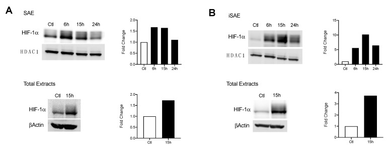
Figure 2.
HIF-1α activation in response to RSV infection. Cells were infected with RSV and harvested at the indicated time points. Nuclear and total lysates of (A) SAE and (B) immortalized SAE (iSAE) cells were then subjected to Western blot analysis to determine HIF-1α levels. Western blot analyses shown here represent one independent experiment. All experiments were repeated at least twice. Densitometric analysis of band intensity, normalized to HDAC-1 for nuclear extracts and βActin for total lysates, is shown to the right of each blot.

