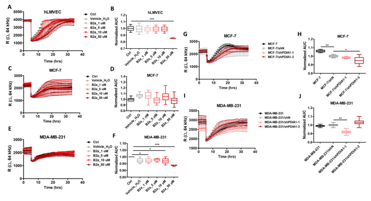
Figure 2.
Effects of PDIA1 inhibition on wound-healing and migration of breast cancer cells and endothelial cells. ECIS Wound-Healing Assay of hLMVEC, MCF-7 and MDA-MB-231 cells and breast cancer sublines with silencing of PDIA1 (shN, shPDIA1-1 and shPDIA1-3). Real time tracings in hLMVECs (A), MCF-7 (C) and MDA-MB-231 (E) cell lines after addition of bepristat 2a at various concentrations (1, 10, 30 or 50 µM) and after PDIA1 silencing in MCF-7 (G) and MDA-MB-231 (I) cell lines. Area under the curve boxplots represent AUC quantitation of changes in migration rate of bepristat 2a-treated hLMVECs (B), MCF-7 (D) and MDA-MB-231 (F) cell lines versus non-treated controls as well as MCF-7 (H) or MDA-MB-231 (J) sublines transduced against PDIA1 (shPDIA1-1, shPDIA1-3) or wild type cells regarding to negative sequence (shN). The line graphs and AUC boxplots represent mean ± SD of three independent experiments. Statistical analysis was calculated using parametric one-way ANOVA followed by Dunnett’s multiple comparisons test (* p = 0.05, ** p = 0.01, *** p = 0.001).

