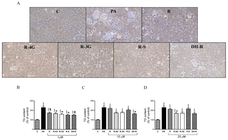
Figure 1.
Optical microscopy images showing structural features at 1 µM (A) and triglyceride content in AML12 hepatocytes exposed to 0.3 M palmitic acid (PA) with or without resveratrol (R) or its metabolites (R-4G, R-3G, R-S and DH-R) at 1 µM (B), 10 µM (C) or 25 µM (D) for 18 h. Data are means ± SEM. *** p < 0.001 vs. control group. # p < 0.05 and ## p < 0.01 vs. PA group.

