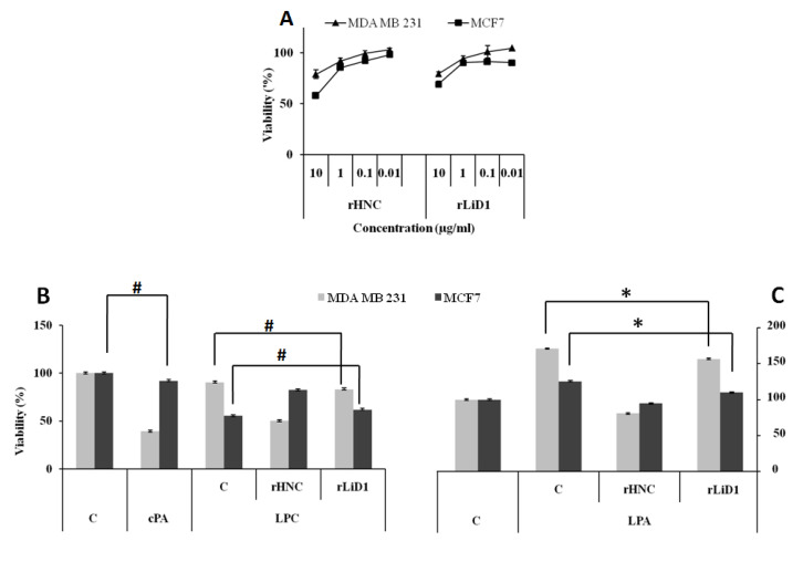
Figure 7.
Cytotoxicity and viability of the MDA-MB-231 and MCF7 cell lines under rHNC and rLiD1 treatment with and without exogenous phospholipids. (A) MDA-MB-231 and MCF7 cells were incubated for 24 h with different concentrations of rHNC and rLiD1 toxins. The cells were detached from the plates and their numbers were assessed by trypan blue counts. (B) The cells were incubated for 24 h with 5 µM LPC alone or added at 1 µg/mL of each toxin. (C) The cells were incubated in the presence of cPA (5 µg/mL) or LPA (5 µM) alone or added to the toxins (1 µg/mL). Viability was measured by the MTT reduction assay. Results were expressed as percentage of mean of four independent experiments and shown as mean of triplicate ± SEM, standard errors of the mean. (*) indicate values statistically different from the control (p < 0.0005), and (#) indicate no significant difference.

