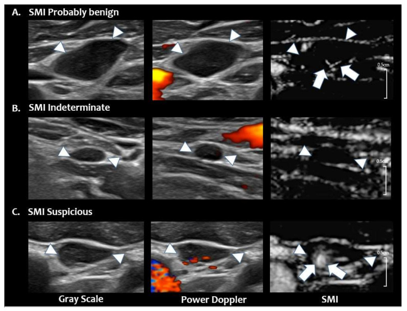
Figure 4.
Reclassified LN categories of SMI. (A) SMI probably benign LN. Grey scale and PDUS images show a hypoechoic LN with loss of hilum and hilar vascularity in the left neck level 3. On SMI, central hilar vascularity is noted (arrows), and this LN is classified as ‘SMI probably benign’ (final diagnosis-benign on FNA-Tg) (B) SMI indeterminate LN. Grey scale and PDUS show a small LN in right neck level 4 with loss of hilum and hilar vascularity. On SMI, no additional vascular signals are noted; therefore, this LN is classified as ‘SMI indeterminate’ (final diagnosis-benign on FNA-Tg). (C) SMI suspicious LN. Grey scale and PDUS show a hypoechoic LN with eccentric cortical thickening and displaced hilar vascularity in left neck level 3. On SMI, abnormal peripheral vascularity is noted at the medial portion of the LN (arrows, final diagnosis-metastatic PTC on CNB). The arrowheads in figures A, B, C indicates the margin of the LN. PTC = papillary thyroid carcinoma, US = ultrasound, LN = lymph node, PDUS = power Doppler ultrasound, SMI = superb microvascular imaging, FNA = fine needle aspiration, CNB = core needle biopsy, Tg = thyroglobulin.

