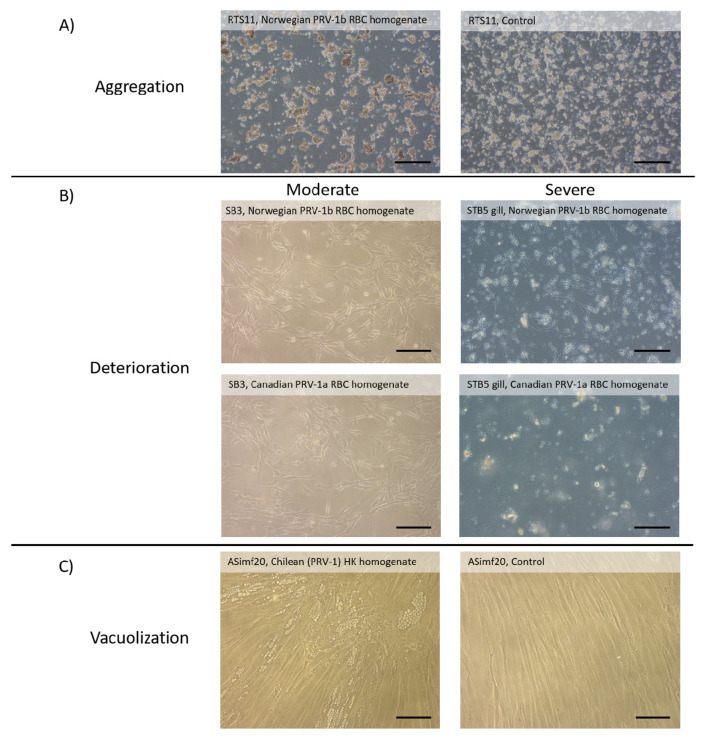
Figure 2.
Rare changes in culture appearance after exposure to PRV-1 homogenates. Although for the vast majority of cultures no change was seen as viewed by phase contrast microscopy, occasionally changes were noted with some cell lines and homogenates. (A) Aggregation of the monocyte/macrophage RTS11 cells over 2 weeks in control cultures (right photo) and the slightly more pronounced aggregation after exposure to PRV-1 RBC homogenates from Norwegian fish (photo on left). (B) Moderate (left side photos) and severe (right side photos) deterioration in the monolayer of cell lines from the sturgeon brain (SB3) and threespined stickleback gill (STB5 gill) two weeks after exposure to either Norwegian PRV-1b (top photos) or Canadian PRV-1a (bottom photos) RBC homogenates. (C) Appearance of vacuoles in cultures of the Atlantic salmon intestinal myofibroblast cell line ASimf20 eight weeks after exposure to AS PRV-1 head kidney homogenates from Chile (left side photo) but not in control cultures (right side photo). Scale bar represents 200 microns.

