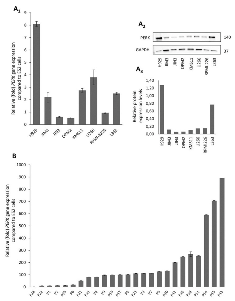
Figure 1.
Protein kinase R (PKR)-like ER kinase (PERK) mRNA (A1) and protein (A2,A3) expression levels in multiple myeloma (MM) cell lines; the uncropped Western Blot figure is shown in Figure S4A1, A2. (B) PERK mRNA expression in isolated CD138+ cells from selected MM patients (n = 25), as determined by Q-RT-PCR. Probing with glyceraldehyde 3-phosphate dehydrogenase (GAPDH) was used as total protein loading reference, whereas β-ACTIN gene expression was used as reference for RNA input. In graphs, means ± SDs from two replicates are shown.

