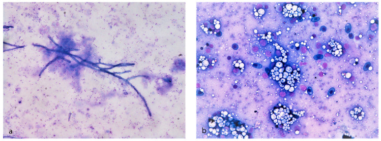
Figure 3.

Cytological examination of Huso huso through May–Grünwald–Giemsa staining. (a) Cytologic smears from coelomic serohemorrhagic exudate showing several rod-shaped blue bacteria in the background and dark-blue branched septate hyphae. (b) Cytologic smears from kidney showing acellular, crystalline, material consistent with mineral deposit (nephrocalcinosis).
