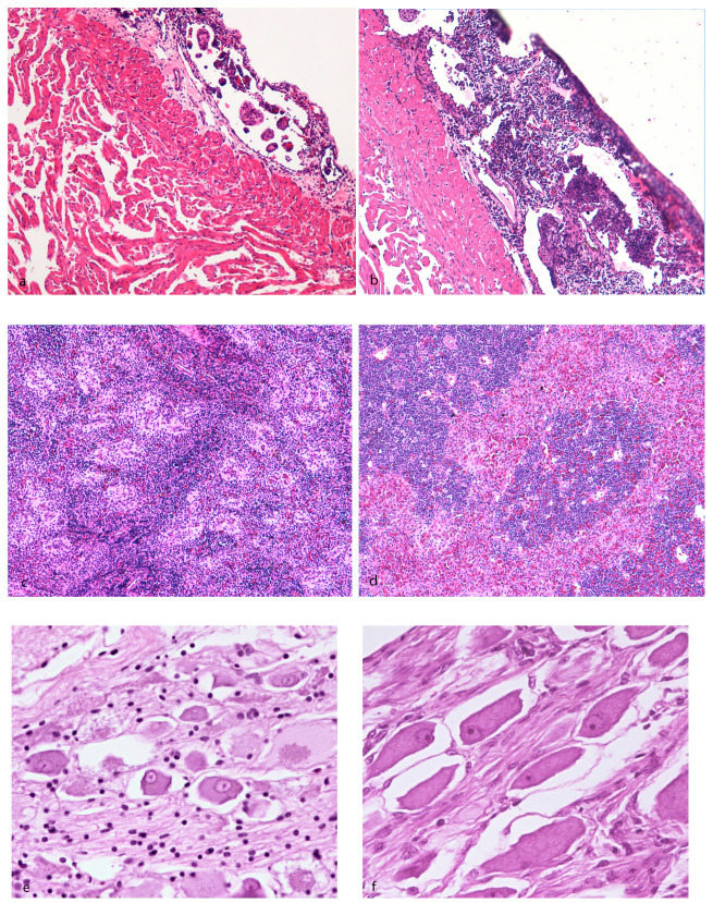
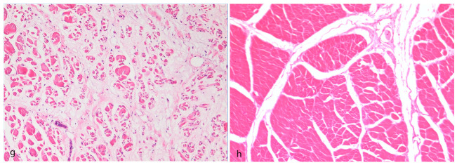
Figure 4.
Hematoxylin–eosin staining of Huso huso. (a) Diseased animal showing the depletion of pericardial–epicardial lymphoid-like tissue compared to (b) aged-matched subject showing normal lymphoid tissue. (c) Diseased animal showing the depletion of lymphoid periarteriolar tissue and thickening of vessel wall (hyalinosis) compared to (d) aged-matched subject showing normal architecture. (e) Diseased animal showing the degeneration/atrophy of pyrenophores in the vicinity of epaxial muscles compared to (f) an aged-matched subject showing normal pyrenophores. (g) Diseased animal showing the degeneration and severe atrophy of skeletal myofibers and perimysial and endomysial edema compared to (h) an aged-matched subject showing normal skeletal muscle.


