Figure 7.
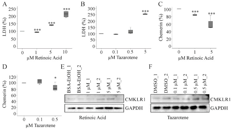
Effect of retinoic acid and tazarotene on chemerin and CMKLR1. (a) HepG2 cells were incubated with increasing concentrations of retinoic acid for 24 h and LDH was measured in the supernatants (n = 4); (b) HepG2 cells were incubated with increasing concentrations of tazarotene for 24 h and LDH was measured in the supernatants (n = 4); (c) HepG2 cells were incubated with retinoic acid (1 and 5 µM) for 24 h and chemerin was measured in the supernatants (n = 4); (d) HepG2 cells were incubated with tazarotene (0.1 and 0.5 µM) for 24 h and chemerin was measured in the supernatants (n = 4); (e) HepG2 cells were incubated with retinoic acid (1 and 5 µM) for 24 h and CMKLR1 was analyzed by immunoblot (n = 2); (f) HepG2 cells were incubated with tazarotene (0.1 and 0.5 µM) for 24 h and CMKLR1 was analyzed by immunoblot (n = 2). Bovine serum albumin (BSA) dissolved in EtOH and dimethylsulfoxid (DMSO) served as solvent controls. * p < 0.05, *** p < 0.001. Statistical test used: Paired t-test.

