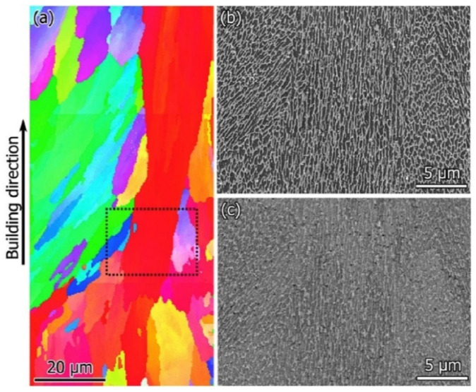
Figure 40.
SEM images of AlSi10Mg SLM samples along the YZ plane: (a) electron back scattered diffraction, (b) secondary electron image and (c) back-scattered image. The inset in (a) shows the area in the electron back-scattered diffraction image, where the secondary and back-scattered images are taken [172]. Copyright 2016. Adapted with permission from Elsevier Science Ltd. under the license number 4803740596529 (Figure 1 [172]), dated 7 April 2020.

