Figure 1.

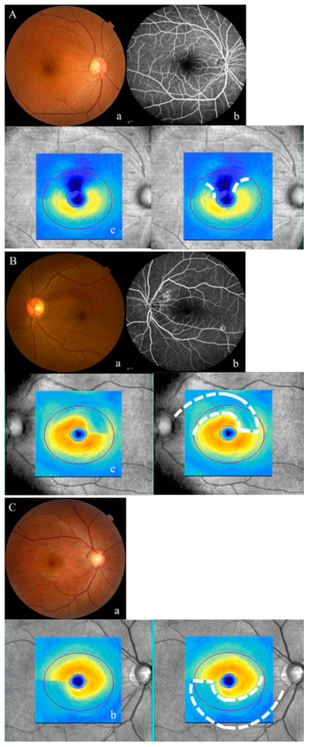
Representative cases from each patient group: patients (A) and (B) have retinal vein occlusion (RVO) and patient (C) has primary open-angle glaucoma (POAG). In patient (A), a fundus photograph (a) shows a superotemporal retinal nerve fiber layer (RNFL) defect. Fluorescein angiography (b) shows superiorly localized abnormal retinal vessels and small fovea non-perfusion. The thickness map of the ganglion cell analysis (GCA) (c) shows non-arcuate and interrupted ganglion cell-inner plexiform layer (GCIPL) thinning with blue/black color in the superior hemisphere. In patient (B), a superotemporal RNFL defect (a) and corresponding regularly arcuate GCIPL thinning on the thickness map (c) are shown. Mild leakage of the fluorescein from an abnormal retinal vessel localized in the superotemporal peripapillary area is shown (b). In patient (C), an inferotemporal RNFL defect (a) and corresponding regularly arcuate GCIPL thinning (b) are shown.
