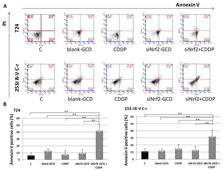
Figure 7.
Apoptosis in T24, 253J B-V C-r cells untreated (C, control) or treated with 8 µg/ml blank-GCD, 2.5 µg/mL CDDP, siNrf2-GCD (0.08 µM siNrf2 in 8 µg/mL GCD), and siNrf2-GCD in combination with CDDP. Apoptosis was checked at 24 h via cytofluorimetric analysis of annexin V/PI stained cells. (A): Flow cytometry profiles of a representative experiment in annexin V/IP stained T24 and 253J B-V C-r cells at 24 h are shown. (B): Quantification of annexin V positive cells in T24 and 253J B-V C-r cells. Results are expressed as percent of the relative control values and are the mean ± standard deviation of three separate experiments. ** p < 0.01.

