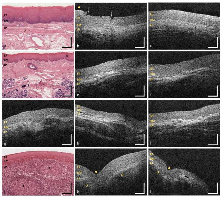
Figure 10.
OCT images of the hard palate (MP12) (b,c), the soft palate (MP13) (e,f), the uvula (MP14) (g), the oropharynx (MP15) (h,i) and the palatine tonsil (MP16) (k,l). Exemplary HE stained histological cross sections depicting the hard palate (a), the soft palate (d) and the palatine tonsil (j) ([44] modified). EP: epithelium, BM: basement membrane, LP: lamina propria, LF: lymphoid follicle; Arrows: epithelial alteration, Yellow dots: palatal ridges and tonsillar crypts. Scale bars: 200 .

