Figure 3.
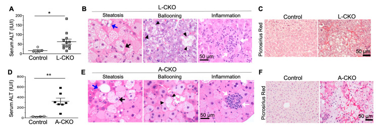
Nonalcoholic steatohepatitis (NASH) features and fibrosis in chow-fed L-CKO mice and A-CKO mice. (A) Serum alanine aminotransferase (ALT) activity in L-CKO mice at 12–18 months of age (n = 5–12 mice per group). * p < 0.05, by Student’s t test. (B) Representative photomicrographs of H&E-stained liver sections from L-CKO mice at 18 months of age; scale bar: 50 µm. Examples of steatosis, hepatocyte ballooning, and inflammation are shown. Macrovesicular steatosis is indicated by blue arrows and microvesicular steatosis is indicated by black arrows. Arrowheads indicate hepatocytes with ballooning degeneration. White arrows indicate inflammatory cells. (C) Picrosirius Red-stained liver sections showing increased fibrosis in L-CKO mice at 18 months of age. (D) Serum ALT activity in A-CKO mice at 6 months of age (n = 4–7 mice per group). ** p < 0.01, by Student’s t test. (E) Representative H&E-stained sections of livers from A-CKO mice at 6 months of age. Examples of steatosis, hepatocyte ballooning, and inflammation are shown; Macrovesicular steatosis is indicated by blue arrows and microvesicular steatosis indicated by black arrows. Arrow heads indicate hepatocytes with ballooning degeneration. White arrows indicate inflammatory cells. (F) Picrosirius Red-stained liver sections showing increased fibrosis in A-CKO mice at 6 months of age. (Reproduced with permission from Shin et al., 2019 [24]).

