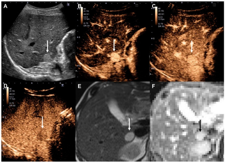
Figure 3.
High-flow hemangioma in a 48-year-old woman with chronic hepatitis B. (A) Gray-scale ultrasound shows a clear hyperechoic mass (arrow) in the liver. (B) The nodule (arrow) shows hyperenhancement at 22 s in the arterial phase of Contrast-Enhanced Ultrasound (CEUS). (C) Enhancement persists at 51 s in the portal venous phase (arrow). (D) In the Kupffer phase, the nodule appears as a slightly hyperechoic mass (arrow) in the liver. (E) Unenhanced T2-weighted MRI shows a clear hyperintense mass (arrow) in the liver. (F) The apparent diffusion coefficient (ADC) map shows a hyperintense mass (black arrow), indicating lower diffusion restriction.

