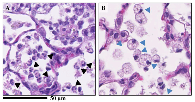
Figure 4.
Lung samples sectioned with H&E staining exposed to nanomaterials intratracheally. (A) the NiO-high dose exposed group; (B) the CeO2-high dose group. There were differences of infiltrating inflammatory cells between nanomaterials. While mainly neutrophils and macrophages were found in the alveoli in the NiO-high dose group (A), macrophage-based inflammatory cell infiltration was observed in the CeO2-high dose group (B). Black arrow heads indicate neutrophils and blue arrow heads indicates macrophages.

