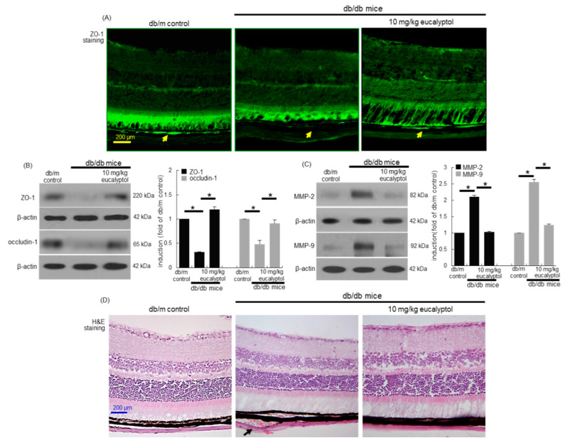
Figure 2.
Immunohistochemical data (A) and western blot data (B and C) showing eye tissue levels of ZO-1, occludin-1, MMP-2 and MMP-9 in eucalyptol-supplemented db/db mice. For the immunohistochemical analysis of ZO-1, green FITC-conjugated secondary antibody was used for visualizing ZO-1 induction (A). Yellow arrows indicate retinal pigment epithelial ZO-1. Tissue extracts were subject to western blot analysis with a primary antibody against ZO-1, occludin-1, MMP-2, MMP-9 and β-actin for an internal control (B and C). The bar graphs (mean ± SEM, n = 3) represent quantitative results of blots in the panels. * Values in respective bar graphs indicate a significant difference at p < 0.05. Inhibition of retinal pigment epithelial detachment from Bruch’s membrane by eucalyptol in db/db mice (D, black arrow). Histological sections of mouse retina were H&E-stained. Each microphotograph is representative of four mice (A and D). Scale bar: 200 μm.

