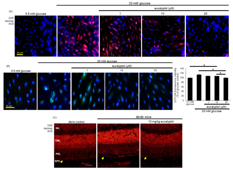
Figure 5.
Inhibition of ROS production by eucalyptol in RPE cells (A and B) and in retina (C). For the measurement of ROS production, the DHE staining was conducted in RPE cells (A) and in retina (C). INL, inner nuclear layer; ONL, outer nuclear layer; PRL, photo receptor layer; RPE, retinal pigment epithelium monolayer. The yellow arrows indicate RPE layer. Furthermore, the DCF-DA staining for ROS production (B) were carried out in eucalyptol-treated RPE cells, and the DCF staining intensity (mean ± SEM, n = 3) was measured. Nuclear staining was done with 4′,6-diamidino-2-phenylindole. DHE- or DCF-stained RPE cells or retina were visualized under fluorescent microscopy. Scale bars: 50–200 μm. * Bar graph values indicate a significant difference at p < 0.05.

