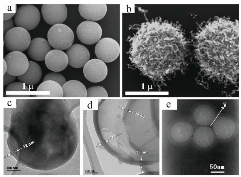
Figure 4.
Scanning electron microscope (SEM) images of (a) SiO2/IO microspheres and (b) “Medusa-like” SiO2/IO/carbon nanofibers and tubes particles [35]. Reproduced with permission from [On Mero et al.], [Langmuir]; published by [American Chemical Society], 2014; Transmission electron microscope (TEM) images of the 1 µm Li2S@C core–shell particles (c) before and (d) after dissolving Li2S [36]. Reproduced with permission from [Caiyun Nan et al.], [Journal of the American Chemical Society]; published by [American Chemical Society], 2014; (e) TEM micrographs of four cores over-coating with one shell structure SiC/SiO2 nanoparticles [37]. Reproduced with permission from [LianZhen Cao et al.], [Journal of Alloys and Compounds]; published by [Elsevier], 2010.

