Figure 2.
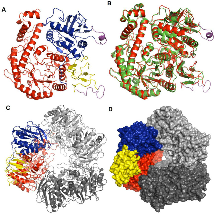
Structure of hla_bga. (A) Overall structure of the hla_bga monomer shown as a ribbon model (domains A, red; B, blue; C, yellow). The modelled loop regions not visible in the electron density are shown in magenta color. (B) Superimposition of hla_bga (red) and tth_bga (green). (C) Trimeric model structure of hla_bga, the domains of one monomer is shown as in Figure 2A while the other two domains are shown in light and dark grey (D) The surface area of trimer structure of hla_bga, one monomer is colored as in Figure 2A while the other two domains are shown in light and dark grey.

