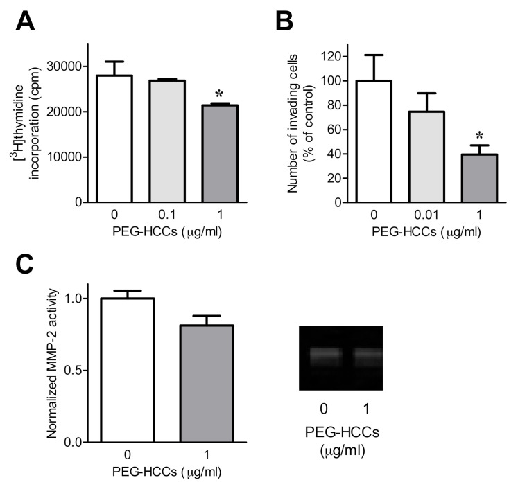
Figure 2.
PEG-HCCs reduce RA-FLS invasion and proliferation. (A) Proliferation of RA-FLS treated with 0, 0.1, or 1 μg/mL PEG-HCCs for 72 h (n = 3). (B) Invasion through Matrigel-coated transwell inserts of RA-FLS in the presence of 0, 0.01, or 1 μg/mL PEG-HCCs (n = 3). Representative images are shown in Supplemental Figure S2. (C) Left, matrix metalloproteinase-2 (MMP-2) secretion of RA-FLS treated with or without 1 μg/mL PEG-HCCs for 24 h (n = 7). Right, example zymography gel of supernatants of RA-FLS treated with or without 1 μg/mL PEG-HCCs for 24 h. * p < 0.05. Data are presented as mean ± SEM.

