Figure 6.
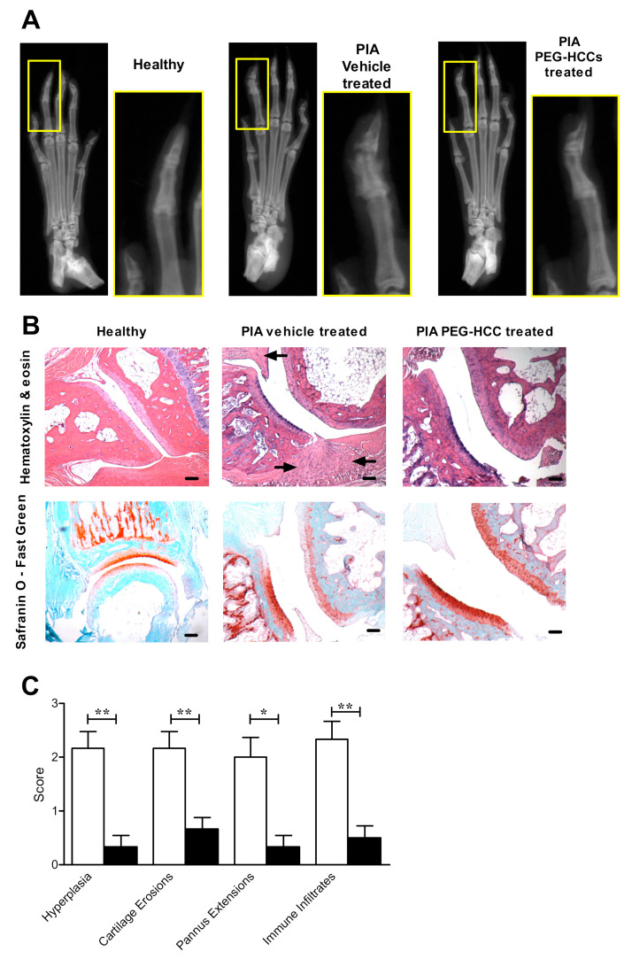
PEG-HCC-treated rats have reduced bone and synovial tissue destruction in PIA. (A) Example X-rays of the hind paws of a healthy rat, a vehicle-treated PIA rat, and a PEG-HCC-treated PIA rat. (B) Example images of hind paw joints of a healthy rat, a vehicle-treated PIA rat, and a PEG-HCC-treated PIA rat, stained with either hematoxylin & eosin (left) or safranin O-fast green (right). Arrows indicate areas of immune infiltrates in the synovium. Scale bar = 100 μm. (C) Quantification of pathologic hallmarks of disease of the joints of PIA rats treated with vehicle (white bars, n = 6) or PEG-HCCs (black bars, n = 6), as determined through analysis of hematoxylin & eosin and safranin O-fast green-stained joint sections. * p < 0.05, ** p < 0.001. Data are presented as mean ± SEM.

