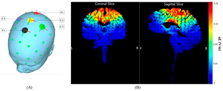
Figure 1.
HD-tDCS electrode placement and estimated current flow within the cortex. (A) The anode (positive electrode) was placed over Cz of the 10/20 EEG template; the four cathodes (negative electrodes) were placed over Fz, C3, Pz, and C4. (B) Coronal and sagittal views of the modeled current flow depicted on a standard brain. Modeling indicated that this induced electric field influenced the foot area of the motor cortex and the primary somatosensory cortex (the area within the white circle). Warmer and cooler colors reflect the larger and smaller modeled electric field normal component (nE, V/m), respectively.

