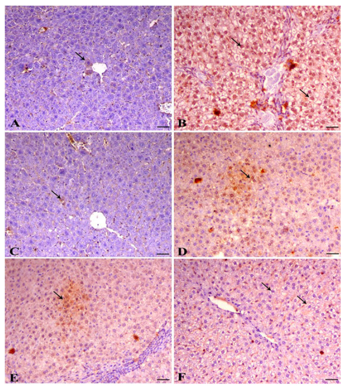
Figure 4.
Immunohistochemical analysis of activated caspase 3 in liver tissue of rats. (A) Liver of control animal showing mild expression of caspase 3 within the hepatocytes (arrow indicates nuclear expression), cleaved form of caspase 3 IHC, bar = 50 µm, ×200. (B) Liver of lead-intoxicated animal showing marked expression of caspase 3 of both cytoplasmic and nuclear expression within the hepatocytes (arrows), cleaved form of caspase 3 IHC, bar = 50 µm, ×200. (C) Liver of normal animal treated with Azolla showing mild expression of caspase 3 within the hepatocytes (arrow indicates cytoplasmic expression), cleaved form of caspase 3 IHC, bar = 50 µm, ×200. (D) Liver of animal treated with both lead and Azolla showing decrease the expression of caspase 3 (arrow), cleaved form of caspase 3 IHC, bar = 50 µm, ×200. (E) Liver of diseased animal pretreated with Azolla showing mild cytoplasmic expression of caspase 3 (arrows), cleaved form of caspase 3 IHC, bar = 50 µm, ×200. (F) Liver of diseased animal treated with Azolla showing marked decrease in caspase 3 expression within the hepatocytes (arrow), cleaved form of caspase 3 IHC, bar = 50 µm, ×200. The labelling indices of caspase 3 were expressed as the percentage of positive cells per total 1000 counted cells in about 10 high-power fields.

