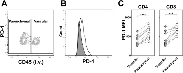
Figure 7.
Increased PD-1 expression associates with cytokine+ T cells from the lung parenchyma. Groups of mice were immunised as per Fig. 1 and cytokine+ CD4 and CD8 T cells derived from the lung parenchyma and lung vasculature compartments detected as described in Figs. 2 and 4. Cells were simultaneously stained for surface expression of PD-1. Representative flow cytometry (A) contoured bivariate plot and (B) histogram both showing the differential expression of PD-1 by cytokine+ CD4 T cells derived from the vascular (CD45+, grey filled line) and parenchymal (CD45-, unfilled line) compartments of the lungs of mice receiving the airway BCG/Ad regimen. (C) Graphs showing the median fluorescence intensity (MFI) of PD-1 staining of cytokine+ CD4 and CD8 T cells derived from the two lung compartments of mice receiving this vaccine regimen. Circles represent individual animals (n = 11). ***p < 0.001 ****p < 0.0001, two-tailed paired t-test. Data representative of one experiment.

