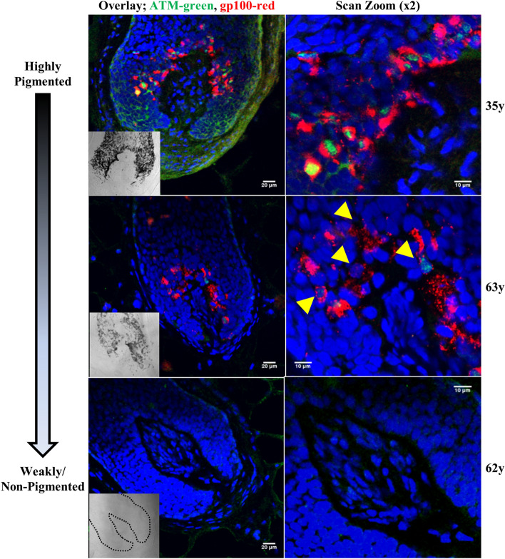
Figure 2.
ATM is expressed within hair follicle bulbar melanocytes and correlates with pigmentation status. Overlays of ATM (green channel) and melanocyte marker GP100 (red channel) in adult human scalp showing strong and exclusive ATM expression in melanocytes within a pigmented follicle (top, 35-year old), less frequent ATM expression in a hair follicle with reduced pigmentation or mild canities (middle, 63-year old), and absent ATM expression in a grey (full canities) hair follicle (lower; 62-year old). Right panels show increased magnification (× 2) of the hair bulb melanocytes present in the central column. Yellow arrows indicate melanocytes with reduced or absent ATM expression in canities-affected scalp follicles. DAPI nuclear stain is shown in blue channel. Scale bar is shown on each image. Brightfield images of follicle pigmentation are shown in the inset image (follicle outline is indicated with black dashed line in 62-year old with full canities). Donor age is indicated on the right.

