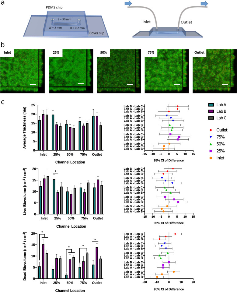
Fig. 3. The publicly available mold design for the microfluidic flow chamber allows reproducible biofilm formation as confirmed by an inter-laboratory comparison.
a Schematic and dimensions of the flow chamber. b Representative images of 72 h MPAO1 WT biofilms grown on the PDMS surface of the device under laminar flow conditions at five different locations along the channel. Biofilms were treated with live/dead staining (green – live cells stained with Syto9; red – dead cells stained with propidium iodide). Scale bar in confocal XY plane: 40 µm. Sagittal XZ section represents biofilm thickness. c COMSTAT data for average thickness, and live/dead biovolume of 72 h MPAO1 WT biofilms generated by three different laboratories, with 95% confidence interval comparisons (3 biological repeats comprising 3 technical repeats per site, i.e., n = 9 biological/n = 27 technical repeats overall; error bars - standard error of mean; 2-way ANOVA with lab and channel location as variables followed by multiple comparisons Tukey test). *p value < 0.05.

