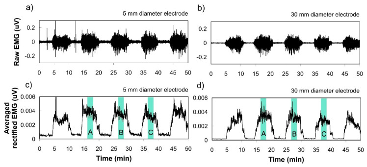
Figure 3.
Examples of SEMG recordings from one subject during intermittent muscle contraction measured by 5 mm and 30 mm diameter electrodes. (a,b) Show an example of raw EMG signals obtained via electrode diameter Ø 5 mm and Ø 30 mm, respectively. (b) clearly shows lower baseline EMG than (a); (c,d) show filtered (20–500 Hz) in analog and full-wave rectified EMG to analyze the amplitude. Three contractions among five, except the first and last trials, were used to calculate the average activated EMG for comparison.

