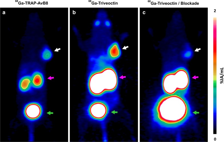
Fig. 6.
Representative PET images (maximum intensity projections, 60 min p.i., OSEM3D reconstruction) of a SCID mouse bearing a subcutaneous MeWo xenograft (human melanoma, positions indicated by white arrow), using 68Ga-TRAP-AvB8 (a 12 MBq, 35 pmol, 350 MBq/nmol) and 68Ga-Triveoctin (b 9 MBq, 25 pmol, 350 MBq/nmol). Blockade (c) was done by administration of 60 nmol Triveoctin, 10 min prior to the radiopharmaceutical. Purple and green arrows indicate presence of activity in kidneys and urinary bladder, respectively

