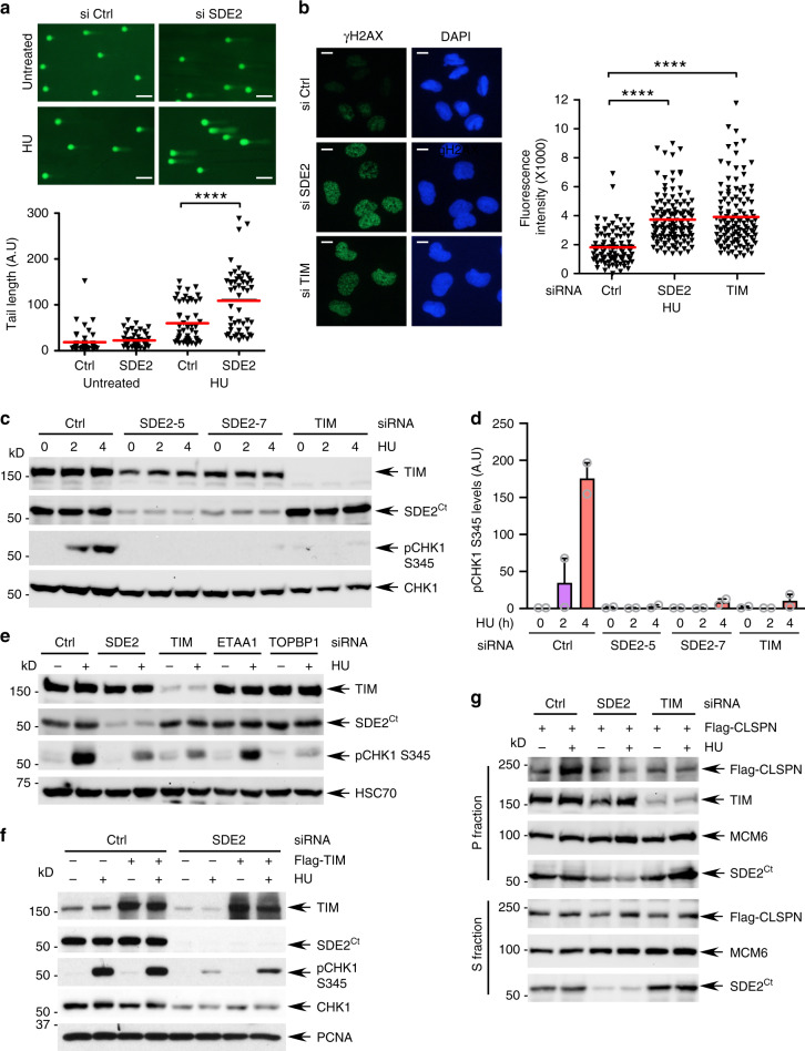
Fig. 5. SDE2 promotes CHK1 activation via TIM and CLSPN.
a Top: representative images of DNA comets after treatment with 2 mM HU for 4 h in siRNA-transfected U2OS cells (n = 2 biologically independent experiments). Scale bar, 50 μm. Bottom: quantification of DNA comet tail lengths. Red bars represent the median (>50 nuclei per sample, ****P < 0.0001, Mann–Whitney test). b Left: representative images of γH2AX staining after treatment with 2 mM HU for 4 h in siRNA-transfected U2OS cells (n = 2 biologically independent experiments). Scale bar, 10 μm. Right: Quantification of γH2AX intensity. Red bars represent the median (>100 nuclei per sample, ****P < 0.0001, Mann–Whitney test). c CHK1 phosphorylation after treatment of 2 mM HU for the indicated times in siRNA-transfected U2OS cells. d Quantification of pCHK1 levels normalized by total CHK1. Mean from two biologically independent experiments is shown. e Comparison of pCHK1 induction in U2OS cells transfected with the indicated siRNAs. f Rescue of pCHK1 induction in U2OS cells transfected with SDE2 siRNA followed by Flag-TIM expression. g Impaired Flag-CLSPN recruitment to the chromatin-enriched P fraction in subcellular-fractionated U2OS cells transfected with SDE2 or TIM siRNA, followed by Flag-CLSPN expression.

