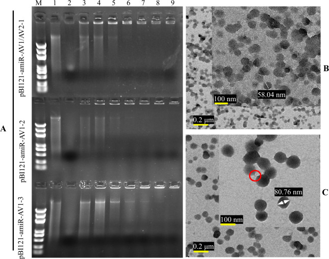
Fig. 1. Characterization of layered double hydroxide (LDH) nanosheets and loaded pDNA.
a pDNA at pDNA:LDH mass ratios of 1:1, 1:2, 1:4, 1:8, 1:10, 1:12, and 1:16, corresponding to lanes 3, 4, 5, 6, 7, 8, and 9, respectively. pDNA only (lane 1) and LDH only (lane 2). M = 5 kb DNA ladder, LDH-bound pDNA was detected by fluorescence in wells. Complete loading was achieved at pDNA–LDH mass ratios of 1:10 (lane 7) and 1:12 (lane 8). b Transmission electron microscopy (TEM) images of the LDH structure. c TEM images of the pDNA–LDH nanocomposite. The surface of the pDNA–LDH composites was smooth, and the nanoparticles tended to have a round shape. The red circle shows a part of the structure of pDNA between LDH nanosheets. Dose ratio = 1:1, T = 298 K

