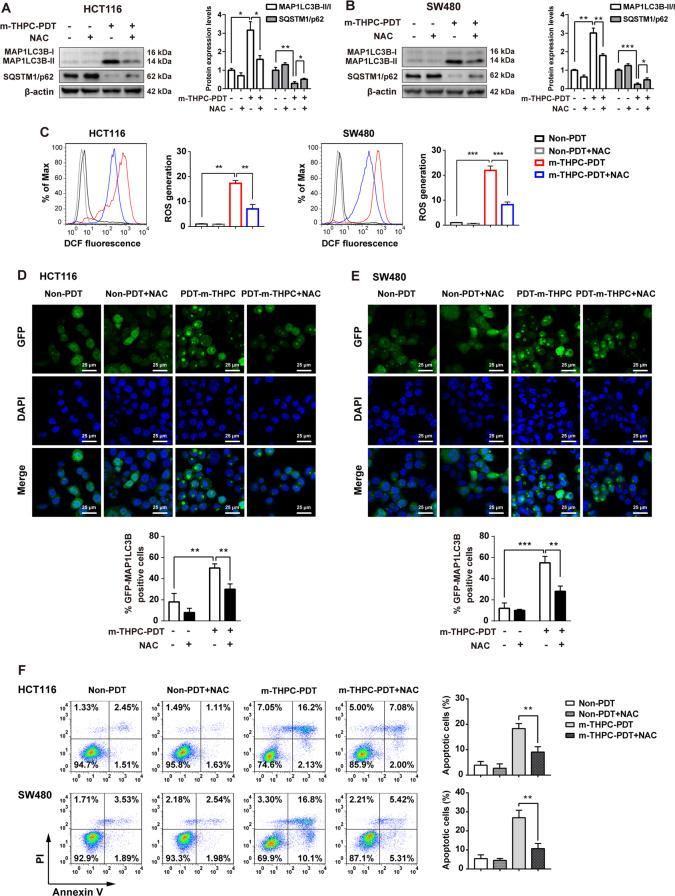
Fig. 4. NAC treatment decreased the autophagy and apoptosis induced by m-THPC-PDT in CRC cells.
HCT116 and SW480 cells were treated with 0.7 μmol/L m-THPC in the presence or absence of 5 mmol/L NAC. NAC and m-THPC were added to the medium 2 and 8 h before irradiation, respectively, and then irradiated with a light dose of 3 J/cm2. Followed by incubation without irradiation for 8 h, A, B Western blot analysis of MAP1LC3B-I, MAP1LC3B-II, and SQSTM1/p62 levels in HCT116 and SW480 cells. C The level of ROS production in HCT116 and SW480 cells was analyzed by flow cytometry to detect DCF fluorescence intensity. D, E GFP-MAP1LC3B puncta were observed by immunofluorescence using a laser scanning confocal microscope. F After 24 h of radiation, flow cytometry was performed to measure cell apoptosis with Annexin V-FITC and PI staining. All data are presented as the mean ± SD, n = 3. **P < 0.01, ***P < 0.001.

