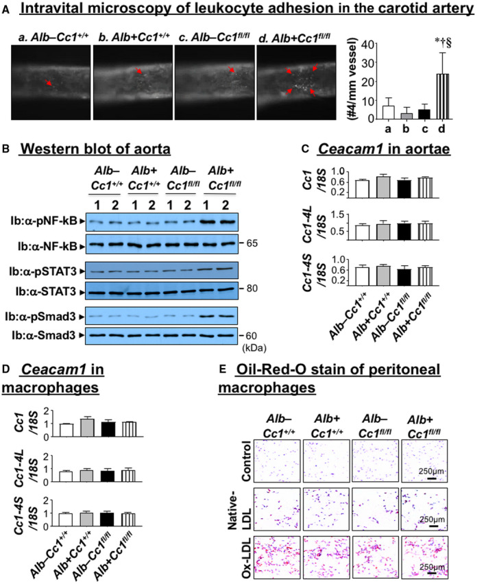
FIG. 6.

Vascular inflammation and leukocyte adhesion. (A) The right jugular vein and left carotid artery were exposed through a middle incision. Carotid arteries were isolated from the surrounding tissues and intravital microscopy of leukocyte adhesion on the carotid artery was assessed in controls (Alb–Cc1+/+, Alb+Cc1+/+, Alb–Cc1fl/fl) and Alb+Cc1fl/fl mutants fed HC for 3 months (n > 5/genotype). Cells that adhered to the vessel wall without rolling or moving for at least 3 seconds were counted over the vessel observed by using an intravital microscope. Video images were analyzed offline for leukocyte adhesion. Total numbers were used for statistical analysis. Values are expressed as mean ± SEM. *P < 0.05 vs. Alb–Cc1+/+ (white); † P < 0.05 vs. Alb+Cc1+/+ (gray); § P < 0.05 vs. Alb–Cc1fl/fl (black). (B) Western analysis was performed on aorta lysates by immunoblotting with antibody (α‐) against phosphorylated NF‐kB, STAT3, and Smad3 antibodies normalized against parallel gels immunoblotted with antibodies against total NF‐kB, STAT3, and Smad3, respectively. Gels represent analysis on 2 mice/group performed on different sets of mice/protein. (C) qRT‐PCR analysis of total, long isoform (Cc1‐4L), and short isoform (Cc1‐4S) of Ceacam1 mRNA levels performed in triplicate relative to 18S (n = 5/genotype). Values are expressed as mean ± SEM. *P < 0.05 vs. Alb–Cc1+/+ (white); † P < 0.05 vs. Alb+Cc1+/+ (gray); § P < 0.05 vs. Alb–Cc1fl/fl (black). (D) Bone marrow macrophages were isolated from tibia and femur of Alb–Cc1+/+ (white), Alb+Cc1+/+ (gray), Alb–Cc1fl/fl (black), and Alb+Cc1fl/fl (hatched) mice and grown in RPMI media supplemented with recombinant M‐CSF. Cells were analyzed by qRT‐PCR in triplicate to assess total Ceacam1, Cc1‐4L, and Cc1‐4S isoforms against 18S. Values are expressed as mean ± SEM. (E) Mice (n = 5/group) were fed HC for 2 months and then injected with thioglycollate into the peritoneal cavity. Their peritoneal macrophages were isolated and cultured in RPMI media and treated in vitro with native LDL (100 µg/mL) or ox‐LDL (100 µg/mL). Cells were fixed with 10% formalin and stained with filtered ORO and counterstained with hematoxylin. Images were taken at 20× magnification. Abbreviation: M‐CSF, macrophage colony‐stimulating factor.
