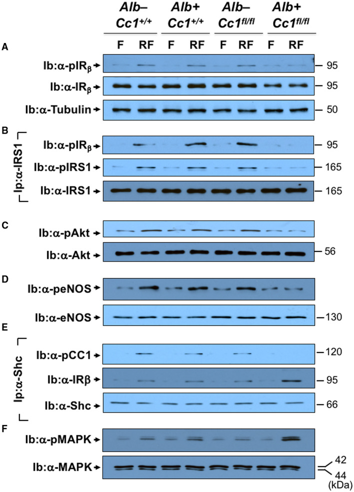
FIG. 7.

Insulin signaling in aortae. Aortae were removed from F and RF mice (n > 6/genotype/treatment) fed HC for 2 months. Western blot analysis was carried out to assess (A) IRβ phosphorylation (α‐pIRβ), normalized to loaded protein IRβ levels, which was in turn normalized by immunoblotting parallel gels with α‐tubulin. (B) Aliquots were subjected to immunoprecipitation with α‐IRS1 antibody followed by immunoblotting with α‐pIRS1 antibody (top gel), normalized to total α‐IRS1 (lower gel). The immunopellet was also immunoblotted with α‐pIRβ antibody to detect binding between IRS1 and IRβ (middle gel). (C) Western analysis was performed by immunoblotting with α‐pAkt and (D) α‐peNOS in parallel to immunoblotting with antibodies against Akt and eNOS, respectively, for normalization. (E) Coimmunoprecipitation was carried out to detect pCC1 (top gel) or IRβ (middle gel) in the Shc immunopellet, as in (B). (F) Immunoblotting with α‐pMAPK in parallel to MAPK for normalization. The apparent molecular weight (kDa) is indicated on the right side of each gel. Analysis was performed on two different mice/genotype using different sets of mice/protein.
