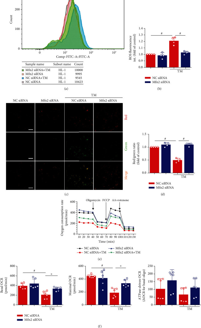
Figure 5.

Decreasing ER-mitochondria interactions by genetic downregulation of mitofusin-2 (Mfn2) protects HL-1 cells from mitochondria dysfunction. (a) Flow cytometry detected HL-1 cells that stained with DCFH-DA. (b) Quantification of ROS by DCFH-DA intensity in (a). Data represent the mean ± SEM (n = 4 independent experiments). (c, d) JC-1 staining. The ratio of red/green fluorescence reflects changes in the mitochondrial membrane potential of HL-1 cells. Data represent the mean ± SEM (n = 4 independent experiments). Scale bar, 100 μm. (e, f) Analysis of oxygen consumption rate (OCR) from a Seahorse XF 24 Extracellular Flux Analyzer. Oligomycin inhibits ATP synthase, FCCP uncouples oxygen consumption from ATP production, and AA+Rotenone inhibits complexes I and III, respectively. Data represent the mean ± SEM (n = 6 independent experiments), #p < 0.01.
