
Figure 2.
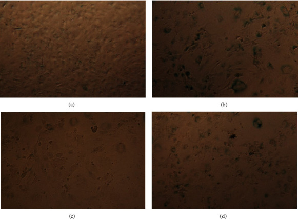
Beta galactosidase activity staining (blue) in 3T3-L1 preadipocytes. Untreated confluent 3T3-L1 preadipocytes show minor amount of senescence cells (a). After BrdU treatment for 8 days, cells show a typical senescence phenotype and increased beta galactosidase activity, which is indicated by blue staining (b). After induction of senescence with BrdU for 8 days, following a treatment with roxithromycin 100 μM for 96 h beta gallactosidase activity decreased resulting in significant less blue stained cells (c). EGCG 100 μM treatment for 96 h after 8days of BrdU treatment could decrease β-gal activity but not in the extend as roxithromycin (d).