Fig. 6.
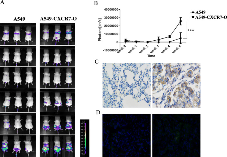
Overexpression of CXCR7 promotes growth of lung cancer cells and metastases in SCID/Beige mice. Mice were injected intravenously with lung cancer cells of A549-GFPL or A549-GFPL -CXCR7-O cells via tail vein injection. a. Continuous images are presented from firefly luciferase bioluminescence imaging for tumors every week until 5 weeks (From top to bottom). Scale bar depicts range of photon flux values as pseudocolor display with red and blue representing high and low values, respectively. b. Quantified data for photon flux from each group of mice. Graph displays mean values + SEM. ***, p < 0.001. c. Lung tissues from A549-GFPL (left) and A549-GFPL-CXCR7-O (right) cell-derived tumors were subjected to immunohistochemical staining for CXCR7.Nuclei were counterstained with hematoxylin (blue) (Magnification:200×). d. GFP fluorescence image. DAPI was used for nuclear detection (Magnification:200×)

