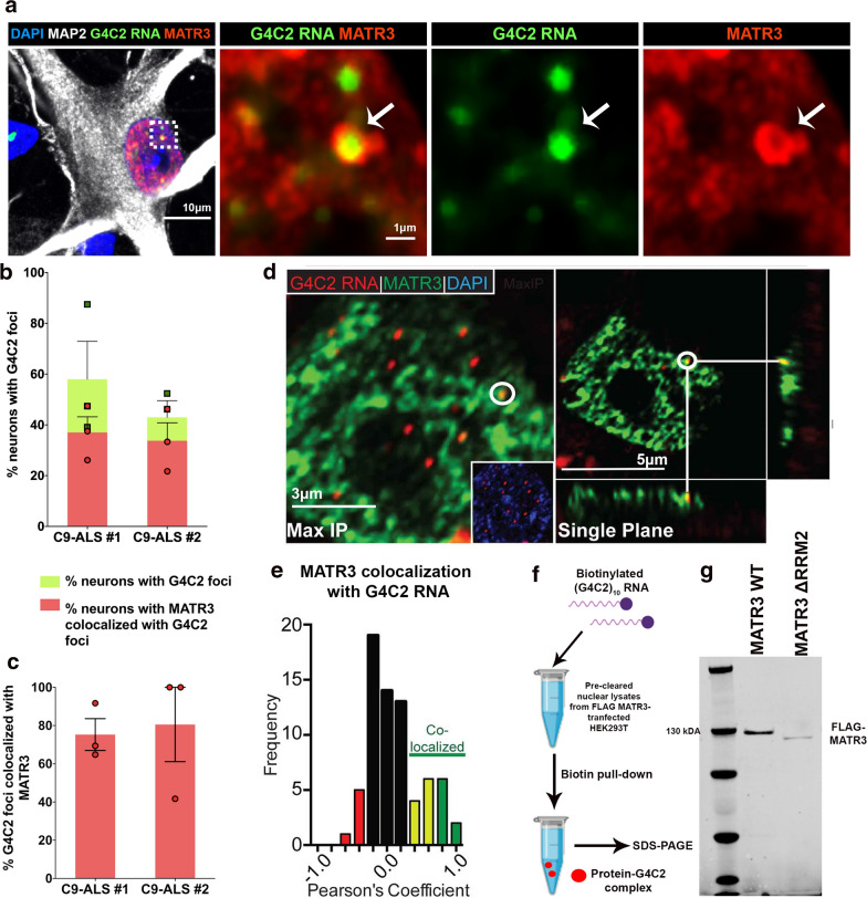
Fig. 1.
MATR3 colocalizes with pathogenic G4C2 RNA foci in C9-ALS iPSC-derived neurons and in post-mortem brain tissue. a Representative confocal image of colocalization (white arrows) between G4C2 RNA foci (green) and MATR3 protein (red) in C9-ALS patient-derived iPSCs differentiated into motor neurons (iPSC-MN), indicated by MAP2 staining (grey). Dotted-white box represents a single G4C2 foci that colocalized with MATR3 and represented in high magnification images to the right. b Quantification of percentage neurons that are positive for G4C2 foci (green bars) in two independent C9-ALS patient iPSC-MNs. Average % neurons that are positive for G4C2 foci in C9-ALS #1 = 58% and in C9-ALS #2 = 48%. Among them, over half of the neurons also showed co-localization between MATR3 and G4C2 RNA foci (red bars). n = 3 differentiations. c Quantification of percentage G4C2 foci per neuron that colocalized with MATR3. Average % G4C2 foci that colocalized with MATR3 in C9-ALS #1 = 75% and in C9-ALS #2 = 80%. n = 3 differentiations. d Representative images of immunohistochemical analysis of MATR3 signal (green) in motor cortex neurons from and C9-ALS patient tissue. RNA FISH analysis performed to examine co-localization between G4C2 RNA foci (red) and MATR3 (green) in nuclei of C9-ALS patient tissue cells. Maximum intensity projection (left) and single plane (right) representative images are shown. Inset within image on left represents overlay between Dapi (blue) and G4C2 foci (red). e Moderate (yellow) to strong (green) Pearson’s coefficients indicate co-localization between RNA foci and MATR3 signal. n = 70 G4C2 RNA foci from 3 C9-ALS patient tissues. f Diagrammatic representation of (G4C2) × 10 pull-down assay. Nuclear lysates from HEK293T cells transiently transfected with either FLAG-MATR3 or FLAG-MATR3-ΔRRM2 were incubated with biotinylated G4C210 RNA. The protein-G4C2 RNA complex was pulled down with streptavidin and then separated by SDS-PAGE. g Immunoblot of biotin-G4C210 pull-down fraction probed for FLAG-MATR3 showed physical interaction between MATR3 and G4C2 RNA. The interaction was moderately diminished between MATR3-ΔRRM2 and G4C2 RNA. Error bars indicate S.E.M

